Mediworks Dry Eye Slit Lamp Diagnostic System

MediworksSKU: DER321

Description


Easy Pathogenic Diagnosis, Providing guidance for customized treatment.
Dry eye diagnostic system


Due to various causes of Dry  Eye Disease,traditional examination  is difficult  to find out the cause and quantify for the treatment.
MediWorks Dry Eye Diagnostic System can provide standardized examination and quantified causes evaluation for  Dry Eye Disease.


Dry eye classification from the 2007 DEWS Report


Non-Invasive Break Up Time


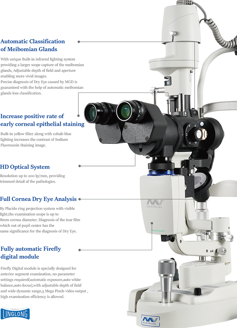
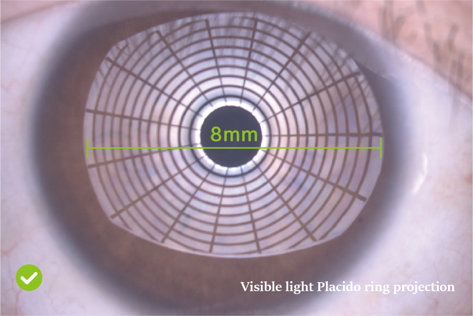
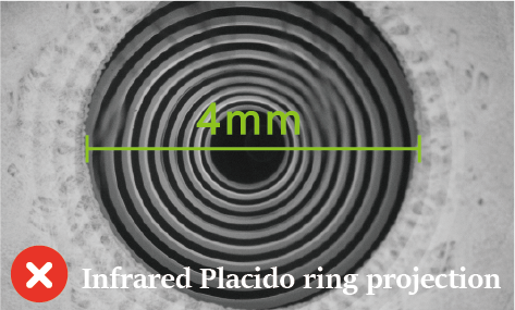
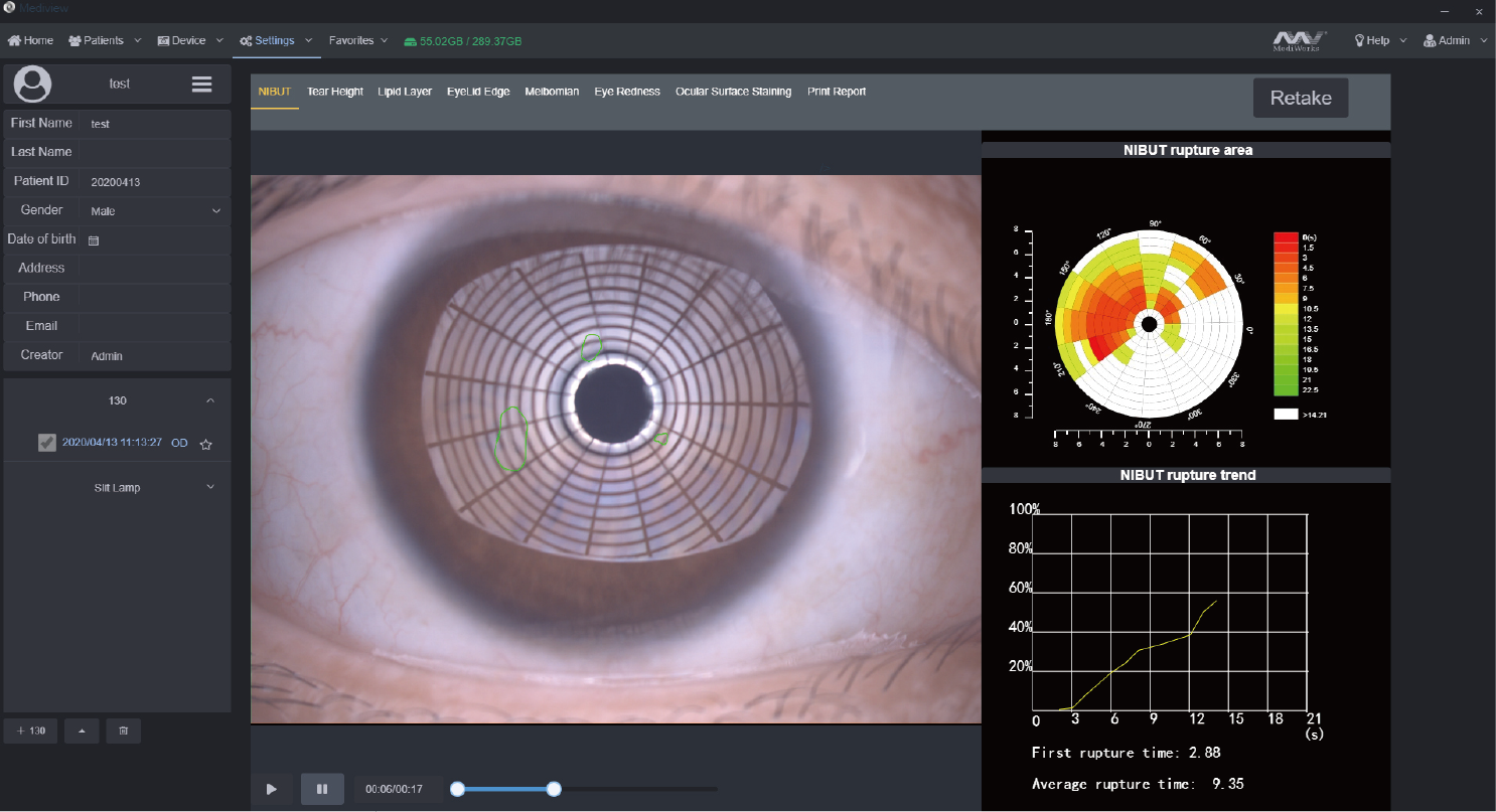
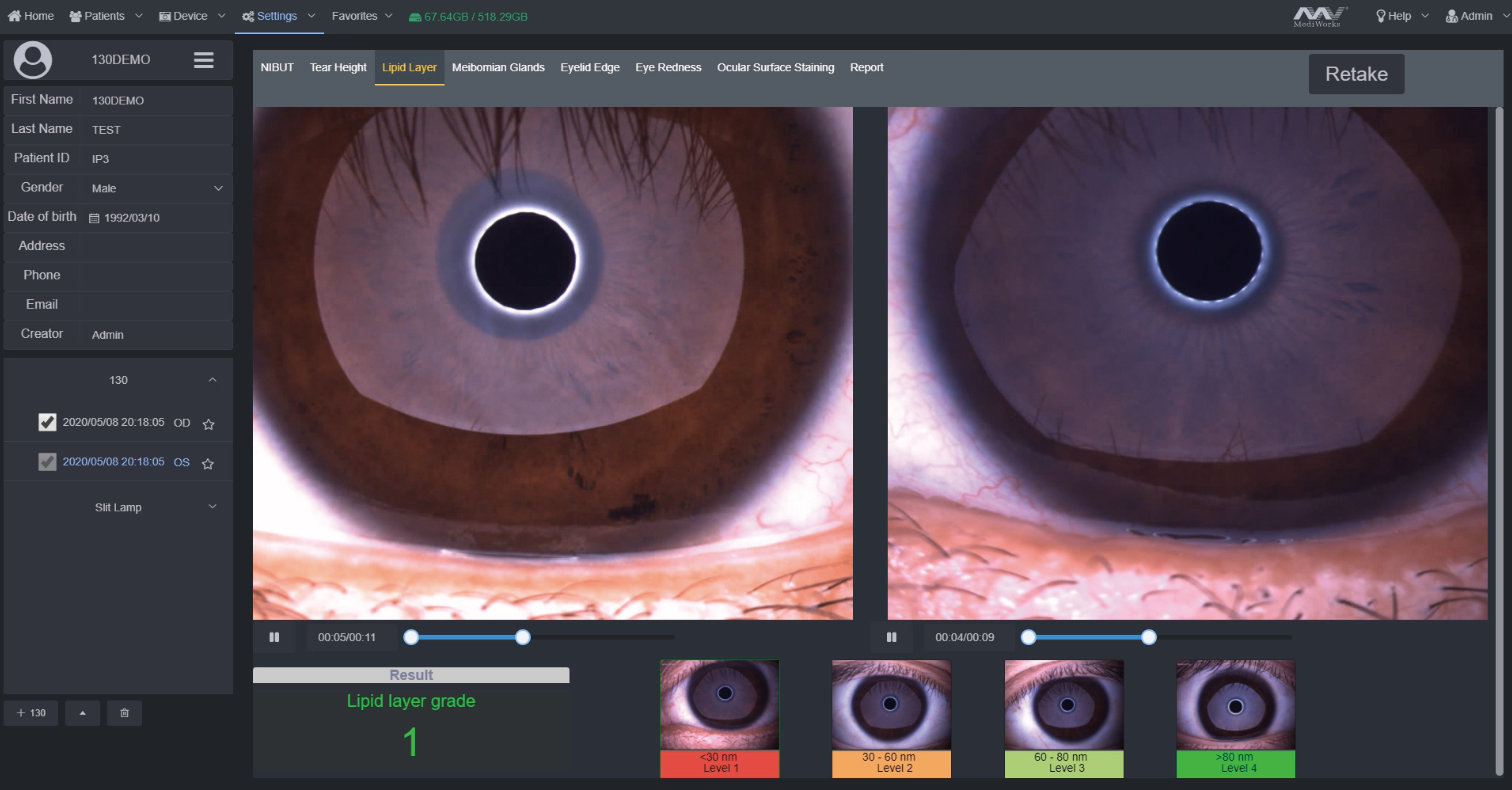
Grade 0 Normal, First Rupture Time: 10 s
Average Rupture Time: 14 s
Grade 1 Warning, First Rupture Time: 6-9 s
Average Rupture Time: 7-13 s
Grade 2 Dry eye, First Rupture Time: 5 s
Average Rupture Time: 7 s


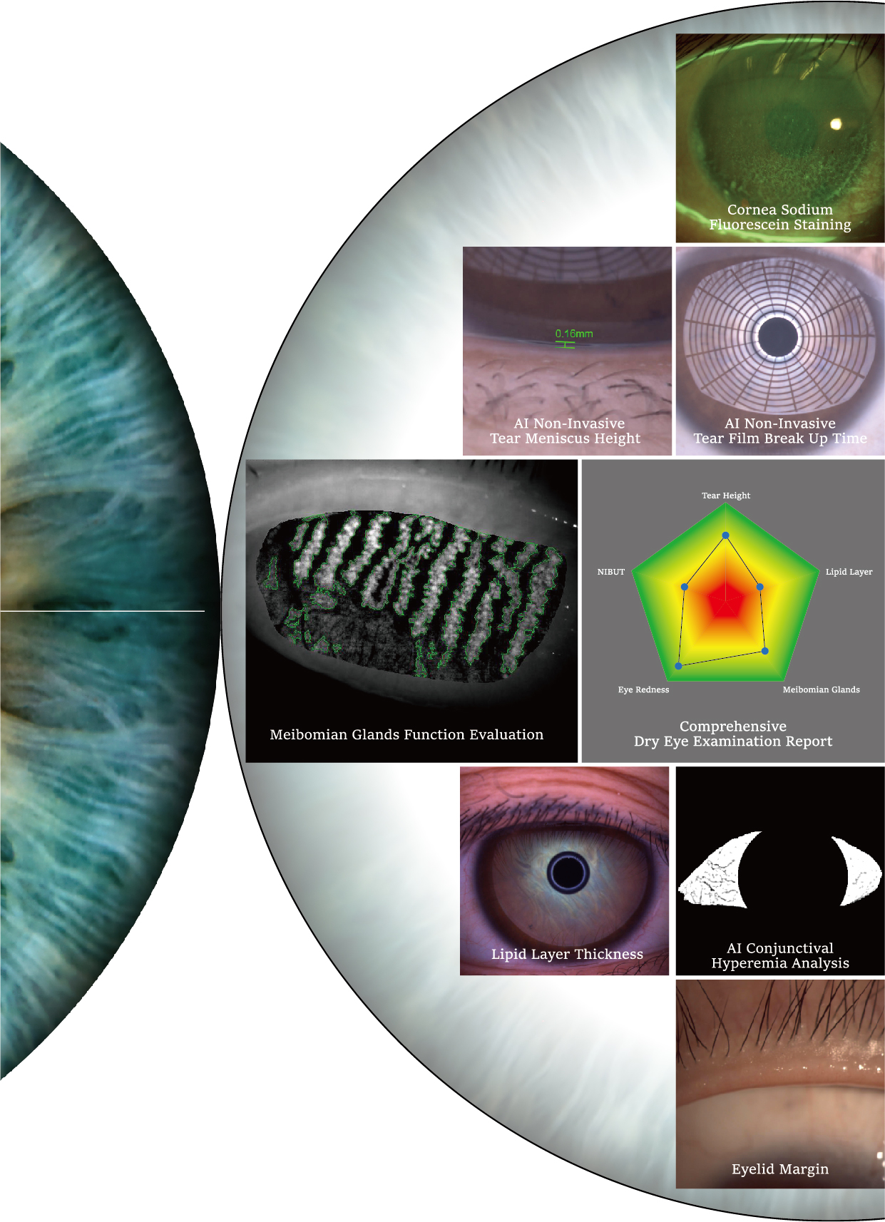
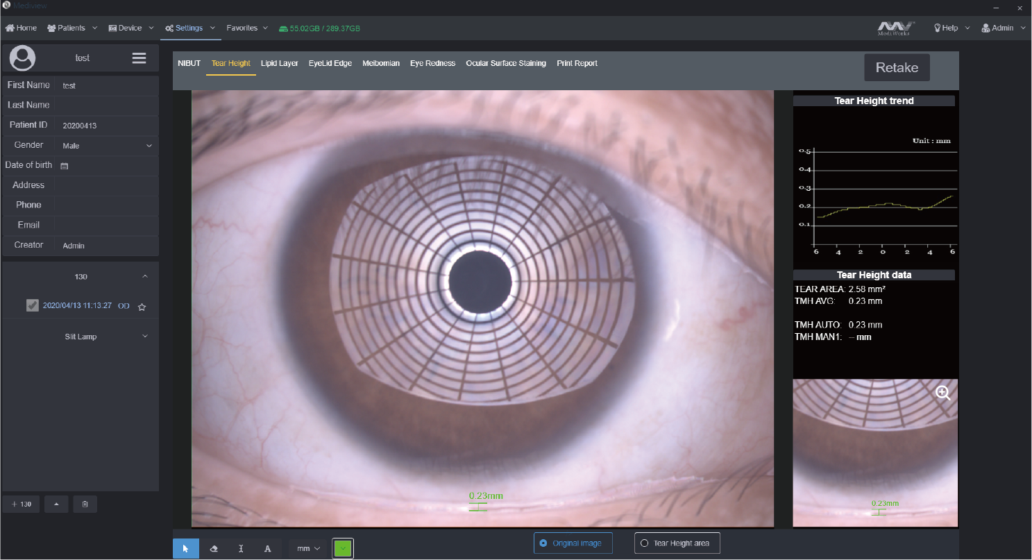
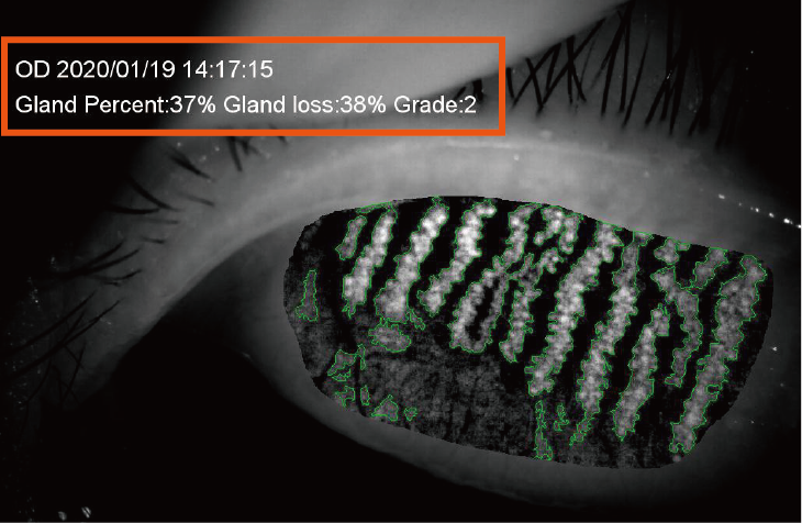
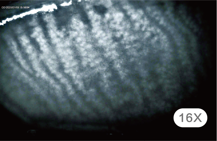
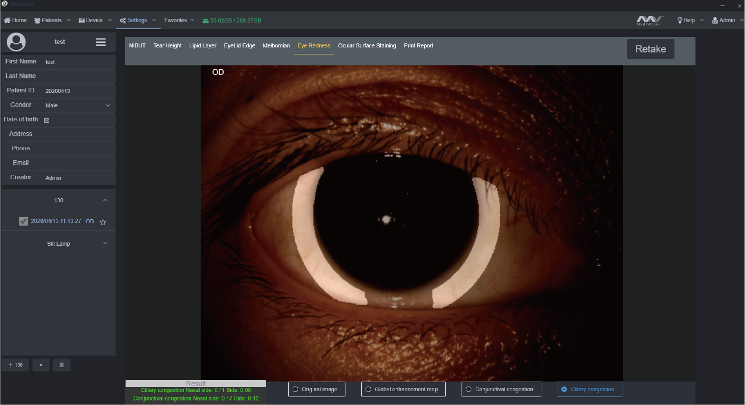
Taking one video brings out AI analysis of NIBUT and Tear Meniscus Height
AI identify the break-up area and analyze NIBUT. Fully Automatic analysis system provides efficient quantified evaluation for the overall stability of the tear film.It automatically acquires the first break up time, average break up time, break up distribution,break up area percentage curve and time distribution.


MediWorks adopts Placido ring projection system with visible light to do the examination,the examination scope  is up to 10mm cornea diameter which brings much more comprehensive diagnosis outcome.
The non-invasive examination avoids the stimulation brought by the traditional Sodium Fluorescein Staining of the Cornea.


Non-Invasive Tear Meniscus Height


Normal: ≥0.2mm
Measures the Tear Meniscus Height and evaluates tear secretion amount and continuity, more efficient and less irritating compared with the traditional Schirmer’s test.


Insufficient tear secretion


Abnormal dynamics and conjunctival chalasis


Evaluation of Meibomian Glands Function


Grade 0: No Meibomian Glands Loss
Grade 1: Meibomian Glands Loss < 1/3 Grade 2: Meibomian Glands Loss 1/3-2/3 Grade 3: Meibomian Glands Loss >2/3

Automatic classification system provides precise and quantified diagnosis of DES caused by meibomian glands dysfunction.
With built-in infrared lighting system,doctors can observe larger image scope of the Meibomian Glands.
Adjustable depth of field makes the glands more prominent and distinguishable against the background.


Meibomian glands loss

Image of Meibomian Glands under high-magnification


Lipid Layer Thickness


(Unit:nm)

Grade 1: <30
Grade 2: 30-60
Grade 3: 60-80
Grade 4: >80

White ring projection system ensures a larger examination area compared to Placido ring.By comparing with the standard grading template and recording the Lipid Layer thickness, it is helpful for judging MGD.


Eyelid Margin 


1. Normal including (Ophthalmic embolism bright, transparent)
2. Mild including (gland cap crown - glandular prominent)
3. Moderate including (glandular fat plug - disappearance of the marginal mucosa, hyperkeratosis)
4. Severe including (uneven margins, disappearance of the meibomian glands - posterior margin Blunt round, thickening, new blood)

MediWorks professional design of optical  system is capable of providing HD digital image that remains clear and sharp even zoom in,  meets the examination  requirements of the overall shape of eyelid margin and its slight change.

AI Analysis of Conjunctival Hyperemia


Normal: ≤2     Abnormal: >2

The unique AI identification system can identify and calculate percentages of conjunctival congestion and ciliary congestions and evaluate severity of eye congestion.

Cornea Sodium Fluorescein Staining


Effectively increases positive rate of early corneal epithelial staining.
Built-in yellow filter along with cobalt-blue filter makes the corneal  sodium fluorescein images more clearly.


Convenient Medical Consultation on Dry Eye Syndrome
Dry Eye Comprehensive Evaluation Report


Smart Patient Management system


Comparison of Patient records
smart patient management system supports repeated comparison among medical records to help doctors customized treatment plans and evaluate treatments.


Patient Management system  allows doctors to build and edit medical records and quickly search the patient case by key words. Besides, doctors can note  patients’ situation via the software. With the DICOM-supported system, Mediview is connected with medical systems in hospitals.


Specifications


Microscope
Microscope Type
Galilean Type
Magnification Change
Revolving Drum 5 steps
Total Magnification
6.3X, 10X, 16X, 25X, 40X

Eyepieces
12.5X

Angle between Eyepieces
10°

Pupillary Adjustment
52mm-80mm

Diopter Adjustment
-8D~+8D

Field of View
Ø36.2mm,  Ø22.3mm,  Ø14mm,  Ø8.9mm,  Ø5.7mm


Slit Illumination


Slit Width
0~14mm continuous (slit becomes a circle at 14mm)

Slit Length
1~14mm continuous

Aperture Diameters
Ø14mm, Ø10mm, Ø5mm, Ø3mm, Ø2mm, Ø1mm, Ø0.2mm

Slit Angle
0°~180°

Slit Inclination
5°, 10°, 15°, 20°

Filters
Heat-absorbing filter, ND filter, Red-free filter, Cobalt blue filter,Yellow filter built-in

Lamp
3V LED Module

Luminance
≥150KLX


Power Supply


Packaging
Input Voltage
100V~240V
Dimension                          770mm x 470mm x 570mm(L/W/H)
Input Frequency
50Hz/60Hz
Gross weight                        23kg
Rated current
1.2A
Net weight                         17kg
Output Voltage
3V LED, Fixation 15V


System Specifications


Digital Module
Automatic exposure/ Automatic white balance / Adjustable depth of field and aperture
Image Sensor
1/1.8-inch sensor / 2.4μm pixel / 5.0M Pixels

Photo Resolution
2592 x 1944

Format
JPEG

Video Resolution
2592 x 1944

Frame of Video
25fps

Video Formats
MP4 H.264

Exposure Mode
Automatic exposure

Transmission Interface
USB 3.0 TYPE-C


System Specifications


PC configuration
i5-8500T 8G 1T+128G 2Gdiscrete graphics

Display
1920×1080 23.8inch

PC system
Windows 10


You may also like

Recently viewed